|
Published in the New Britain Herald,
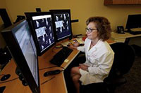
Special to The Press, centralcommunications.com, Sunday, June 14, 2015.  Joel Gelber, MD, is Chief of Radiology at The Hospital of Central Connecticut and Medical Director of the hospital’s Imaging Center, which recently relocated to the new Medical Arts Center facility at 201 North Mountain Road in Plainville. The new center features state-of-the-art imaging technology, including a 3T MRI, which is taking MRI capabilities to a new level. Joel Gelber, MD, is Chief of Radiology at The Hospital of Central Connecticut and Medical Director of the hospital’s Imaging Center, which recently relocated to the new Medical Arts Center facility at 201 North Mountain Road in Plainville. The new center features state-of-the-art imaging technology, including a 3T MRI, which is taking MRI capabilities to a new level.
Q. What is MRI technology? How does it work? Dr. Gelber: Magnetic resonance imaging, or MRI, has become the tool of choice for hospitals and healthcare providers throughout the world in getting the most accurate images of the human anatomy to determine if someone has a wide range of possible health issues like a tumor or a ruptured disk. The technology, which relies on magnetic fields to produce a non-radioactive scan of the body’s interior, has become so popular in recent years that it is believed that there are over 25,000 MRI scanners now in use across the world. Q. What are some of the advantages and shortcomings of traditional MRI technology? Dr. Gelber: MRI offers enormous advantages in terms of providing patients with a quick and accurate diagnosis, as well as providing greater flexibility for a wider range of treatment options. Its impact has been revolutionary, yet until recently it still featured shortcomings such as limited resolutions; an inability to capture high-resolution images of certain body parts such as the prostate without causing patient discomfort; and a small bore – the donut-shaped opening where patients are confined during the exam – that left many patients feeling claustrophobic. Q. Can you describe the new 3T MRI technology and how it is overcoming those issues? Dr. Gelber: It’s only been until very recently that technology has advanced to the point where these issues are no longer a problem. In particular, the Phillips Ingenia 3T MRI system, which is available at The Hospital of Connecticut Imaging Center as well as a handful of other imaging services providers in Connecticut, is proving to be a game-changer. 3T refers to the strength of the magnet in the system, which is the strongest available for clinical use, allowing for higher resolution imaging. Among other things, the 3T offers high-resolution images of the prostate without causing patient discomfort and, with the use of MRI-Ultrasound fusion software, can guide urologists to biopsy tumors that could only be found on an MRI scan. Q. How about the issue with the small bore and the claustrophobic sensation it can cause some patients? Dr. Gelber: While the 3T MRI is expanding our ability to bring improved outcomes to patients, it is also bringing enhanced comfort. The 3T has the widest bore now available on the market, significantly reducing the claustrophobic sensation that many patients have felt with earlier machines.
Joel Gelber, MD, has been practicing in the field of radiology for over 25 years and is Board-Certified in radiological imaging. © Copyright 2015, Central Connecticut Communications, New Britain, CT.
|